
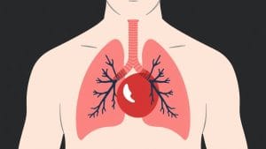
The New England Journal of Medicine has shared medical images of a lung hernia occurring alongside a chest-wall blood collection, flagged under emergency medicine and radiology.
The New England Journal of Medicine posted the case to its social media channels, categorising it under emergency medicine and radiology. The images relate to a lung hernia — a condition where lung tissue pushes through a gap in the chest wall — appearing together with a haematoma (a collection of blood beneath the skin or within tissue) on the chest wall.
Lung hernias are uncommon. They can follow trauma, surgery, or intense physical strain, and are typically diagnosed using imaging such as a CT scan (a detailed X-ray that produces cross-sectional pictures of the body).
The journal did not supply further clinical detail, patient information, or outcome data in the post itself.
—
*For any chest pain, breathing difficulty, or unexplained swelling on the chest wall, seek urgent medical attention. Call NHS 111 for non-emergency guidance, or 999 in an emergency.*
Source: @NEJM
Lung Hernia with Chest-Wall Bleeding: New England Journal Posts Rare Clinical Images Quiz
5 questions